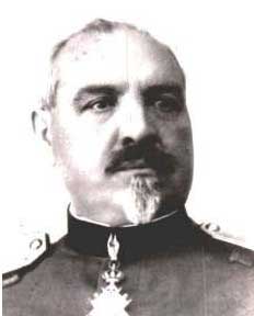
Dr Adolph Hempt, founder and first director

Dr Milan Nikolić
Pasteur Institute was founded in 1921. Founder and first director was
dr Adolf Hempt, whose vaccine (nerve tissue) against rabies was used in Europe since late 80` XX century.
Pasteur Institute in Novi Sad today is National Reference Laboratory for Rabies, which work on rabies diagnosis and prevention for humans and animals, producing and distribution of HRIG and statistics of rabies cases in Serbia. Pasteur Institute is member of RABNET WHO sponsored international network for rabies information.
Pasteur Institute in Novi Sad is EU approved laboratory for FAVN test (Fluorescent Antibody Virus Neutralisation). Test is the method of choice for determining the levels of antibody to rabies virus in pets serum.
HISTORY
The first Pasteur Institute in Serbia, was founded in Nis in 1900. Its great work, mainly on rabies and variola vaccine production was stopped during the First World War: In the health policy of the Monarchy of Serbs, Croatians and Slovenians, the priority was given to the organization of preventive medicine. A great number of bacteriological laboratories, stations for desinfection, antituberculosis, antialchoholic and antitrachoma clinics were founded. Their work was coordinated by Central Hygienic Institute in Belgrade, founded in 1924. The Pasteur Institute in Nis was restored in 1928. and new ones were founded in Novi Sad and Cetinje.
The Pasteur Institute in Novi Sad, which was founded in 1921. became central in the Country in 1928, thanks to a new method of producing inactivated ether-phenol vaccine by Dr Adolph Hempt, the founder and it’s first director. Along with the improvement of human vaccination using his lypovaccine with live fix rabies virus. More than 50 years of permanent work of many generations of doctors and veterinarians, were necessary extinguish urban rabies finally in Yugoslavia. However, in the 70’s, the silvatic rabies began to spread so we have the similar inconvenient situation with permanent appearance of animal rabies in whole country.
Dr Milan Nicolich, Hempt’s successor, was second director of Pasteur Institute which was famous scientist in the Europe. He organized a industrial production of Hempt’s vaccine

The direct fluorescent antibody test (DFA) is most frequently used to routine diagnose of rabies. This test can be performed on brain tissue of animals suspected of being rabid. The rapid and accurate laboratory diagnosis of rabies infections in animals is essential for timely administration of postexposure prophylaxis. Within a few hours, a diagnostic laboratory can determine whether or not an animal is rabid and inform the responsible medical personnel. The laboratory results may save a patient from unnecessary physical and psychological trauma, if the animal is not rabid. All rabies laboratories in the Yugoslavia (3) perform this test on animals suspected of having rabies.
The DFA test is based on the principle that an animal infected by rabies virus will have rabies virus antigen present in its tissue. Because rabies is present in nervous tissue (and not blood like many other viruses) the ideal tissue to test for the presence of rabies antigen is brain. The most important part of a DFA test is flouresecently-labelled anti-rabies antibody. Pasteur Institute in Novi Sad produced fluorescent-labelled anti-rabies antibody on hamsters and goat. When labeled antibody is added to rabies-suspect brain tissue, it will bind to rabies antigen if it is present. Unbound antibody can be washed away and the areas where the antigen has bound antibody will appear as a bright fluorescent apple green color when viewed with a fluorescence microscope. If rabies virus is absent there will be no staining. The rabies antibody in the DFA test is primarily directed against the nucleoprotein (antigen) of the virus. Rabies virus replicates in the cytoplasm of cells, and infected cells may contain large round or oval inclusions containing collections of nucleoprotein (N) or smaller collections of antigen that appear as dust-like fluorescent particles if stained by the DFA procedure.
Several tests are necessary to confirm or rule out rabies in a human intra-vitam. Tests are performed on samples of serum, spinal fluid, skin biopsies from the nape of the neck, and saliva. Routinely, serum and spinal fluid are tested for antibodies to rabies virus. Saliva maybe tested by virus isolation or nested reverse transcription polymerase chain reaction (RT-PCR) methods.
General histopathology
Although it is a nonspecific and less-sensitive method of diagnosing rabies, histological examination of biopsy or autopsy tissues is occasionally useful in diagnosing unsuspected cases of rabies that have not been tested by routine methods. When brain tissue from a rabies virus-infected animal is stained with a histological stain, such as hematoxylin and eosin, a trained microscopist may recognize evidence of encephalomyelitis.
Histopathologic evidence of rabies encephalomyelitis in brain tissue and meninges includes the following:
1. Mononuclear infiltration
2. Perivascular cuffing of lymphocytes or polymorphonuclear cells
3. Lymphocyte foci
4. Babes nodules consisting of glial cells
5. Negri bodies
Negri bodies
Rabies inclusions were detected in 1903. Dr. Adelchi Negri reported that he had identified what he believed was the etiologic agent of rabies, the Negri body. This was a round or oval inclusion body sometimes observed within the cytoplasm of nerve cells of animals infected with rabies. Negri bodies may vary in size from 0.25 to 27 µm. These inclusions are found most frequently in the pyramidal cells of Ammon’s horn, in the Purkinje cells of the cerebellum, and in the cells of the medulla and various ganglia. With Seller’s stain, Negri bodies appear magenta in color and have small (0.2 µm to 0.5 µm), dark-blue interior basophilic granules. The histological technique may detect Negri bodies in only 50% of known positive samples in contrast to the DFA which detects these inclusion bodies in essentially 99% of samples.
Mouse inoculation test
Mouse inoculation test, in spite of its simplicity, depends greatly on the accuracy of its performance for dependable results. White mice of both sexes are equally susceptible to rabies virus. Mice of all ages are susceptible to intracerebral inoculation of rabies virus, but suckling mouse up to 3 days are more sensitive. Single mouse dose for intracerebral inoculation is 0,03 ml 10% suspension of suspected brain. Observation period should be 28 days after inoculation. If mouse showing symptoms of rabies, should be killed and prepared for DFA test.
Other tests for diagnosis of rabies
Other tests for diagnosis and research, such as electron microscopy (EM), immunohistochemistry (IHC), RT-PCR, and isolation in cell culture are useful tools for studying the virus structure, molecular typing, and virulence of rabies viruses.
Amplification methods
When samples contain a small amount of rabies virus, it may be difficult to detect the virus by routine methods. Virus isolation in cell cultures, such as mouse neuroblastoma cells (MNA) or baby hamster kidney (BHK) cells, increases the sensitivity of detection by increasing the virus concentration as virus replicates in the cell cultures. These are excellent methods for confirming the DFA test results.
Another method of amplification of rabies virus uses biochemical methods to produce many copies of the rabies virus nucleic acid. Rabies RNA can be copied into a DNA molecule using reverse transcriptase (RT). The DNA copy of rabies can be amplified using the technique of polymerase chain reaction (PCR). With this method, rabies virus RNA can be enzymatically amplified as DNA copies. Forty cycles of amplification, can produce up to 1012 times the original amount of nucleic acid in the virus sample. This technique is used to confirm DFA results and to detect rabies virus in saliva and skin biopsy samples that contain minute amounts of rabies virus.
Rabies is a preventable viral disease of mammals most often transmitted through the bite of a rabid animal. The vast majority of rabies cases reported in Yugoslavia each year occur in foxes. Domestic animals account for less than 10% of the reported rabies cases, with cats most often reported rabid, and occasionally in dogs, cattle, sheep and goats.
Rabies virus infects the central nervous system, causing fatal encephalitis and ultimately death. Early symptoms of rabies in humans are nonspecific, consisting of fever, headache, and general malaise. As the disease progresses, neurological symptoms appear and may include insomnia, anxiety, confusion, slight or partial paralysis, excitation, hallucinations, agitation, hyper salivation, difficulty swallowing, and hydrophobia. Death usually occurs within days of the onset of symptoms.
Public health importance of rabies
Over the last 100 years, rabies in the Yugoslavia has changed dramatically. More than 90% of all animal cases reported annually now occur in wildlife; In northern part of country urban rabies was eliminated before 1960, in Central Serbia at the end of seventies and in Kosovo and Metohija in early 1980. The principal rabies hosts today are foxes. The latest human deaths in the Yugoslavia were registered in 1980. on Kosovo i Metohija. Modern prophylaxis has proven with 100% successful. In Yugoslavia, animals bite about 10.000 persons annually. Only 10% of them are treated against rabies.
Rabies prevention
Post exposure prophylaxis in Yugoslavia is organized over the Net of Antirabic stations in Institutes of Public Health or Clinics for Infective Diseases across the country and it is free of coast for all of the patients. In last 10 years HRIG is produced in Yugoslavia, with high quality and good potency. Since 1990 the Institute for Blood Transfusion in Belgrade and Pasteur Institute in Novi Sad have been producing HRIG. Nowadays human rabies vaccine from cell culture is imported but in few last years we developed human rabies vaccine on BHK cell culture for human use. Now it is in clinical trails on human volunteers. We have good results in all with high antibodies titer and mild adverse reaction at the inoculation site.
The Virus
Classification
Rabies virus belongs to the order Mononegavirales, viruses with a nonsegmented, negative-stranded RNA genome. Within this group, viruses with a distinct „bullet“ shape are classified in the Rhabdoviridae family, which includes at least three genera of animal viruses, Lyssavirus, Ephemerovirus, and Vesiculovirus. The genus Lyssavirus includes rabies virus, Lagos bat, Mokola virus, Duvenhage virus, European bat virus 1 & 2 and a newly discovered Australian bat virus.
Structure
Rhabdoviruses are approximately 100-300 nm long and 75 nm wide. The rabies genome encodes five proteins: nucleoprotein (N), phosphoprotein (P), matrix protein (M), glycoprotein (G) and polymerase (L). All rhabdoviruses are having two major structural components: a helical ribonucleoprotein core (RNP) and a surrounding envelope. In the RNP, the genome RNA is tightly encased by the nucleoprotein. Two other viral proteins, the phospoprotein and the large protein (or polymerase) are associated with the RNP. The glycoprotein forms approximately 400 trimeric spikes that are tightly arranged on the surface of the virus. The M protein is associated both with the envelope and the RNP and may be the central protein of rhabdovirus assembly. Rabies virions are bullet-shaped with 10-nm spike-like glycoprotein peplomers covering the surface. Rabies has a genome of RNA. The rabies genome contains 5 genes that encode 5 proteins designated as N, P, M, G, and L. The arrangement of these proteins and the RNA genome determine the structure of the rabies virus. The rabies virus genome is single-stranded, antisense, nonsegmented, RNA of approximately 12 kb. There is a leader (LDR) of 50 nucleotides, followed by N, P, M, G, and L genes.
Replication
The fusion of the rabies virus envelope to the host cell membrane (adsorption) initiates the infection process. The interaction of the G protein and specific cell surface receptors may be involved. After adsorption, the virus penetrates the host cell and enters the cytoplasm by pinocytosis. The virions aggregate in the large endosomes (cytoplasmic vesicles). The viral membranes fuse to the endosomal membranes, causing the release of viral RNP into the cytoplasm. Since lyssaviruses have a linear single-stranded, negative-stranded ribonucleic cid (RNA) genome the complementary „positive“ strand of RNA must be transcribed before virus replication. A viral polymerasease transcribes the negative strand of RNA into leader RNA and five capped and polyadenylated mRNAs, which are translated into proteins. Translation, which involves the synthesis of the N, P, M, G and L proteins, occurs on free ribosomes in the cytoplasm. Within the central nervous system (CNS), there is preferential viral budding from plasma membranes. Conversely, virus in the salivary glands buds primarily from the cell membrane into the acinar lumen. Specific viral budding into the saliva coupled with virus-induced behavioral changes, maximizes the chance of viral perpetuation through the directed bite and infection of a new host.
Immunity after vaccination
The history of active immunization by vaccine began 115 years ago with Louis Pasteur, who sought a post exposure vaccination against rabies in man. Pasteur protected a 9-year old boy after a rabid dog bite, using dried neural cord as a vaccine. Since Pasteur, vaccination schemes have been based on a continuous vaccine stimulus given for many days and/or multiple injections on the first day. The firs generation type of vaccine (nerve tissue culture) fails to fulfill the present standards of an efficient and safe immunoprophylaxis due to its low protective efficiency, high rate of neurocomplications and death rate. Current immunoprophylaxis is based on inactivated cell culture vaccine. Early, high, and long-lasting antibody induction became the criterion of a successful vaccination regimen.
Development of WHO approved BHK -21 (baby hamster kidney) cell culture vaccine request investigation immune response and its protective effects in man. Pasteur Institute in Novi Sad was beginning vaccination of human volunteers with this vaccine and our first results are very good.
Form 1 – REQUEST FOR DETERMINATION OF RABIES ANTIBODIES BY FAVN
(Fluorescent Antibody Virus Neutralization) TEST

